Файл:CSF circulation.png
Перейти до навігації
Перейти до пошуку
Розмір при попередньому перегляді: 800 × 350 пікселів. Інші роздільності: 320 × 140 пікселів | 640 × 280 пікселів | 1024 × 448 пікселів | 1956 × 856 пікселів.
Повна роздільність (1956 × 856 пікселів, розмір файлу: 673 КБ, MIME-тип: image/png)
| Відомості про цей файл містяться на Вікісховищі — централізованому сховищі вільних файлів мультимедіа для використання у проектах Фонду Вікімедіа. |
Опис файлу
| Опис |
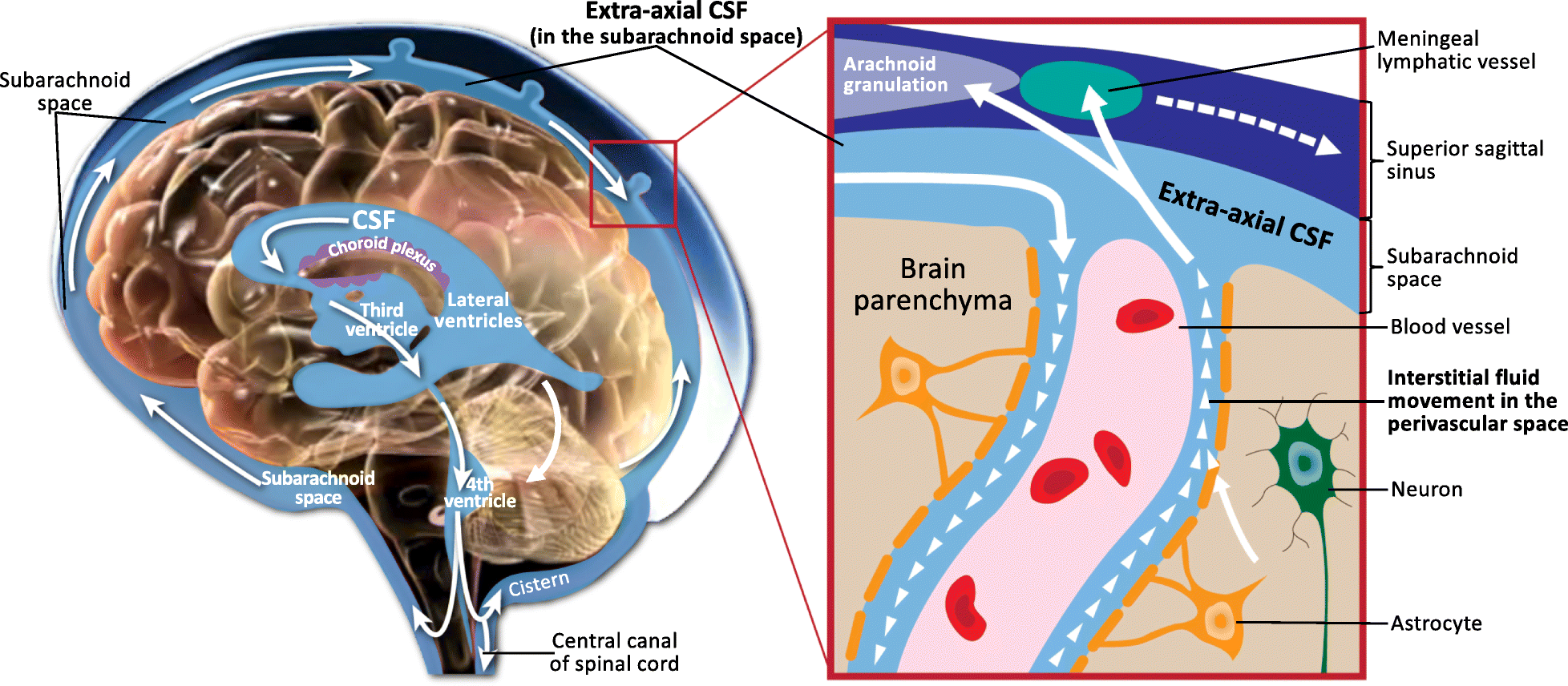
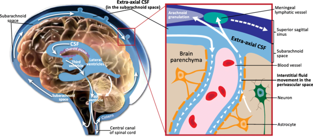
English: Schematic of CSF circulation, CSF outflow systems, and the anatomy of various CSF compartments. CSF is produced by the choroid plexus in the ventricles, where it delivers growth factors to progenitor cells that originate on the surface of the ventricles, and then proliferate into neurons and migrate to form the cerebral cortex. CSF circulates from the lateral, third and fourth ventricles to the cisterns of the brain, and then flows into the subarachnoid space, where it envelops the cortical convexities of the brain (EA-CSF). Inset box: From the subarachnoid space, there is retrograde influx of CSF into the parenchyma, where CSF and interstitial fluid interact in the perivascular space, alongside blood vessels that course throughout the brain. Astrocytes lining the perivascular space aid in transporting fluid that removes inflammatory waste proteins (e.g., Aβ), which are continually secreted by neurons as byproducts of neuronal activity and would otherwise build up in the brain. Finally, fluid carrying these inflammatory waste products returns to the subarachnoid space (EA-CSF) and drains into meningeal lymphatic vessels and arachnoid granulations. |
| Час створення | First Online: 13 December 2018 |
| Джерело | Shen MD. Cerebrospinal fluid and the early brain development of autism. J Neurodev Disord. 2018;10(1):39. Published 2018 Dec 13. https://dx.doi.org/10.1186%2Fs11689-018-9256-7 |
| Автор | Mark D. Shen |
Ліцензування
Цей файл доступний на умовах ліцензії Creative Commons Із зазначенням авторства 4.0 Міжнародна
- Ви можете вільно:
- ділитися – копіювати, поширювати і передавати твір
- модифікувати – переробляти твір
- При дотриманні таких умов:
- зазначення авторства – Ви повинні вказати авторство, надати посилання на ліцензію і вказати, чи якісь зміни було внесено до оригінального твору. Ви можете зробити це в будь-який розсудливий спосіб, але так, щоб він жодним чином не натякав на те, наче ліцензіар підтримує Вас чи Ваш спосіб використання твору.
Історія файлу
Клацніть на дату/час, щоб переглянути, як тоді виглядав файл.
| Дата/час | Мініатюра | Розмір об'єкта | Користувач | Коментар | |
|---|---|---|---|---|---|
| поточний | 11:07, 16 червня 2019 | | 1956 × 856 (673 КБ) | Was a bee | {{Information |Description={{en|1=Schematic of CSF circulation, CSF outflow systems, and the anatomy of various CSF compartments. CSF is produced by the choroid plexus in the ventricles, where it delivers growth factors to progenitor cells that originate on the surface of the ventricles, and then proliferate into neurons and migrate to form the cerebral cortex. CSF circulates from the lateral, third and fourth ventricles to the cisterns of the brain, and then flows into the subarachnoid space... |
Використання файлу
Така сторінка використовує цей файл:
Глобальне використання файлу
Цей файл використовують такі інші вікі:
- Використання в ar.wikipedia.org
- Використання в en.wikipedia.org
- Використання в fr.wikibooks.org
- Використання в ko.wikipedia.org
- Використання в pt.wikipedia.org