Файл:Salivary gland epithelial tissue growing in vitro.jpg
Матеріал з Вікіпедії — вільної енциклопедії.
Перейти до навігації
Перейти до пошуку
Salivary_gland_epithelial_tissue_growing_in_vitro.jpg (542 × 540 пікселів, розмір файлу: 37 КБ, MIME-тип: image/jpeg)
| Відомості про цей файл містяться на Вікісховищі — централізованому сховищі вільних файлів мультимедіа для використання у проектах Фонду Вікімедіа. |
Опис файлу
| ОписSalivary gland epithelial tissue growing in vitro.jpg |
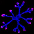
English: We can study how organs grow and regenerate by dissecting out their cellular components and grow them in vitro, in the lab. The picture shows a specific type of cellular tissue from embryonic salivary glands of mouse, called epithelium, that differentiates into the functioning organ (salivary gland) under certain conditions. In the laboratory, we can dissect out the embryonic organs, culture its separated components, and add growth factors to the culture conditions that will simulate normal growth, stimulate cellular growth and tissue regeneration. The picture is a confocal microscopy image showing cells of the epithelium of an embryonic salivary gland (a marker for these cells is used to label them in blue) and the growing cells (labeled in red/pink) that proliferate in the tip of the tissue, which drives its elongation and further formation of ducts and other specialized tissue structures important for functionality of the organ. English: We can study how organs grow and regenerate by dissecting out their cellular components and grow them in vitro, in the lab. The picture shows a specific type of cellular tissue from embryonic salivary glands of mouse, called epithelium, that differentiates into the functioning organ (salivary gland) under certain conditions. In the laboratory, we can dissect out the embryonic organs, culture its separated components, and add growth factors to the culture conditions that will simulate normal growth, stimulate cellular growth and tissue regeneration. The picture is a confocal microscopy image showing cells of the epithelium of an embryonic salivary gland (a marker for these cells is used to label them in blue) and the growing cells (labeled in red/pink) that proliferate in the tip of the tissue, which drives its elongation and further formation of ducts and other specialized tissue structures important for functionality of the organ. |
| Час створення | |
| Джерело | Власна робота |
| Автор | Irebustini |
This picture was taken in a research laboratory in the National Institute of Dental and Craniofacial Research in Bethesda, MD.
Ліцензування
Я, власник авторських прав на цей твір, добровільно публікую його на умовах такої ліцензії:
Цей файл доступний на умовах ліцензії Creative Commons Із зазначенням авторства 4.0 Міжнародна
- Ви можете вільно:
- ділитися – копіювати, поширювати і передавати твір
- модифікувати – переробляти твір
- При дотриманні таких умов:
- зазначення авторства – Ви повинні вказати авторство, надати посилання на ліцензію і вказати, чи якісь зміни було внесено до оригінального твору. Ви можете зробити це в будь-який розсудливий спосіб, але так, щоб він жодним чином не натякав на те, наче ліцензіар підтримує Вас чи Ваш спосіб використання твору.
| Це зображення було завантажено в рамках Конкурсу наукової фотографії 2017. |
Підписи
Додайте однорядкове пояснення, що саме репрезентує цей файл
Об'єкти, показані на цьому файлі
зображує
Якесь значення без елемента на сайті Вікідані
2 січня 2012
Історія файлу
Клацніть на дату/час, щоб переглянути, як тоді виглядав файл.
| Дата/час | Мініатюра | Розмір об'єкта | Користувач | Коментар | |
|---|---|---|---|---|---|
| поточний | 15:04, 3 листопада 2017 |  | 542 × 540 (37 КБ) | Irebustini | User created page with UploadWizard |
Використання файлу
Така сторінка використовує цей файл:
Метадані
Файл містить додаткові дані, які зазвичай додаються цифровими камерами чи сканерами. Якщо файл редагувався після створення, то деякі параметри можуть не відповідати цьому зображенню.
| Програмне забезпечення | Picasa |
|---|---|
| Версія Exif | 2.2 |
| Номер зображення (ID) | d295a60c729b47031002d4dbe39430a7 |

