Файл:Rabies encephalitis Negri bodies PHIL 3377 lores.jpg
Перейти до навігації
Перейти до пошуку

Розмір при попередньому перегляді: 800 × 532 пікселів. Інші роздільності: 320 × 213 пікселів | 640 × 425 пікселів | 1024 × 681 пікселів | 1280 × 851 пікселів | 1801 × 1197 пікселів.
Повна роздільність (1801 × 1197 пікселів, розмір файлу: 438 КБ, MIME-тип: image/jpeg)
| Відомості про цей файл містяться на Вікісховищі — централізованому сховищі вільних файлів мультимедіа для використання у проектах Фонду Вікімедіа. |
Опис файлу
| Опис |
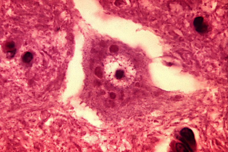
English: This micrograph depicts the histopathologic changes associated with rabies encephalitis prepared using an H&E stain.
Note the Negri bodies, which are cellular inclusions found most frequently in the pyramidal cells of Ammon's horn, and the Purkinje cells of the cerebellum. They are also found in the cells of the medulla and various other ganglia. |
||
| Час створення | |||
| Джерело |
|
||
| Автор | Content Provider(s): CDC/Dr. Daniel P. Perl | ||
| Ліцензія (Повторне використання цього файлу) |
Copyright Restrictions: None – This image is in the public domain and thus free of any copyright restrictions. As a matter of courtesy we request that the content provider be credited and notified in any public or private usage of this image. | ||
| Інші версії |
 |
Ліцензування
Це зображення є роботою працівника Центру контролю та запобігання хвороб (агентство Міністерства охорони здоров'я і соціальних служб США), зробленою чи сфотографованою ним як частина його обов'язків. Як робота Федерального уряду США, це зображення перебуває в суспільному надбанні в США.
|
Історія файлу
Клацніть на дату/час, щоб переглянути, як тоді виглядав файл.
| Дата/час | Мініатюра | Розмір об'єкта | Користувач | Коментар | |
|---|---|---|---|---|---|
| поточний | 07:27, 26 жовтня 2011 | | 1801 × 1197 (438 КБ) | Ghainmem | Higher-resolution version |
| 18:01, 30 травня 2006 |  | 700 × 465 (57 КБ) | Patho | {{Information| |Description=ID#: 3377 Description: This micrograph depicts the histopathologic changes associated with rabies encephalitis prepared using an H&E stain. Note the Negri bodies, which are cellular inclusions found most frequently in the pyra |
Використання файлу
Такі сторінки використовують цей файл:
Глобальне використання файлу
Цей файл використовують такі інші вікі:
- Використання в ar.wikipedia.org
- Використання в de.wikibooks.org
- Використання в fr.wikipedia.org
- Використання в ja.wikipedia.org
- Використання в th.wikipedia.org