Файл:Electron micrograph of neuromuscular junction (cross-section).jpg
Перейти до навігації
Перейти до пошуку
Нема версії з більшою роздільністю.
Electron_micrograph_of_neuromuscular_junction_(cross-section).jpg (433 × 289 пікселів, розмір файлу: 95 КБ, MIME-тип: image/jpeg)
| Відомості про цей файл містяться на Вікісховищі — централізованому сховищі вільних файлів мультимедіа для використання у проектах Фонду Вікімедіа. |
.jpg)
Опис файлу
| Опис |
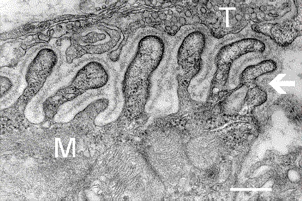
English: Electron micrograph showing a cross-section through the neuromuscular junction. T is the axon terminal, M is the muscle fiber. The arrow shows junctional folds with basal lamina. Postsynaptic densities are visible on the tips between the folds. The scale is 0.3 µm. |
| Час створення | Originally uploaded to en.wikipedia on 10 березня 2006. |
| Джерело | Synapse Web at the National Institute of Mental Health, National Institutes of Health; originally from en.wikipedia; description page is/was here. |
| Автор | National Institute of Mental Health; originally uploaded by Nrets at en.wikipedia. |
Ліцензування
This image is a work of the National Institutes of Health, part of the United States Department of Health and Human Services, taken or made as part of an employee's official duties. As a work of the U.S. federal government, the image is in the public domain.
 |
||
| Цей файл визнано вільним від відомих обмежень з боку закону про авторські права, включаючи всі пов'язані і суміжні права. | ||
Журнал завантажень локального файлу
(All user names refer to en.wikipedia)
- 2006-03-10 20:07 Nrets 433×289×8 (97758 bytes) Electron micrograph showing a cross section through the neuromuscular junction. T is the axon terminal, M is the muscle fiber. The arrow shows junctional folds with basal lamina. Postsynaptic densities are visible on the tips between the folds. Scale is 0
Історія файлу
Клацніть на дату/час, щоб переглянути, як тоді виглядав файл.
| Дата/час | Мініатюра | Розмір об'єкта | Користувач | Коментар | |
|---|---|---|---|---|---|
| поточний | 03:41, 22 березня 2007 | | 433 × 289 (95 КБ) | Fran Rogers | {{Information |Description=Electron micrograph showing a cross section through the neuromuscular junction. T is the axon terminal, M is the muscle fiber. The arrow shows junctional folds with basal lamina. Postsynaptic densities are visible on the tips be |
Використання файлу
Такі сторінки використовують цей файл:
Глобальне використання файлу
Цей файл використовують такі інші вікі:
- Використання в ar.wikipedia.org
- Використання в cs.wikipedia.org
- Використання в de.wikipedia.org
- Використання в es.wikipedia.org
- Використання в fa.wikipedia.org
- Використання в gl.wikipedia.org
- Використання в he.wikipedia.org
- Використання в ko.wikipedia.org
- Використання в pt.wikipedia.org
- Використання в ru.wikipedia.org
- Використання в zh.wikipedia.org
.jpg)