Файл:Free-living amebic infections.png

Матеріал з Вікіпедії — вільної енциклопедії.
Перейти до навігації Перейти до пошуку
Повна роздільність(1365 × 1170 пікселів, розмір файлу: 715 КБ, MIME-тип: image/png)

Wikimedia Commons logo Відомості про цей файл містяться на Вікісховищі — централізованому сховищі вільних файлів мультимедіа для використання у проектах Фонду Вікімедіа.
Це зображення біологічного об'єкта має бути конвертоване у векторний формат SVG. Це дає декілька переваг, докладніше про які Ви можете прочитати на сторінці Commons:Media for cleanup. Якщо Вам вже зараз доступна векторна версія даного зображення, завантажте її, будь ласка. Після завантаження замініть цей шаблон на такий: {{vector version available|Назва_завантаженого_файлу.svg}}.

Опис файлу

Опис
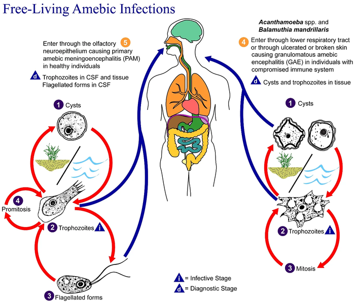
English: This is an illustration of the life cycle of the parasitic agents responsible for causing “free-living” amebic infections.

For a complete description of the life cycle of these parasites, select the link below the image or paste the following address in your address bar: http://www.dpd.cdc.gov/dpdx/HTML/FreeLivingAmebic.htm Free-living amebae belonging to the genera Acanthamoeba, Balamuthia, and Naegleria are important causes of disease in humans and animals. Naegleria fowleri produces an acute, and usually lethal, central nervous system (CNS) disease called primary amebic meningoencephalitis (PAM). N. fowleri has three stages, cysts (1) , trophozoites (2) , and flagellated forms (3) , in its life cycle. The trophozoites replicate by promitosis (nuclear membrane remains intact) (4) . Naegleria fowleri is found in fresh water, soil, thermal discharges of power plants, heated swimming pools, hydrotherapy and medicinal pools, aquariums, and sewage. Trophozoites can turn into temporary flagellated forms which usually revert back to the trophozoite stage. Trophozoites infect humans or animals by entering the olfactory neuroepithelium (5) and reaching the brain. N. fowleri trophozoites are found in cerebrospinal fluid (CSF) and tissue, while flagellated forms are found in CSF.

Acanthamoeba spp. and Balamuthia mandrillaris are opportunistic free-living amebae capable of causing granulomatous amebic encephalitis (GAE) in individuals with compromised immune systems. Acanthamoeba spp. have been found in soil; fresh, brackish, and sea water; sewage; swimming pools; contact lens equipment; medicinal pools; dental treatment units; dialysis machines; heating, ventilating, and air conditioning systems; mammalian cell cultures; vegetables; human nostrils and throats; and human and animal brain, skin, and lung tissues. B. mandrillaris however, has not been isolated from the environment but has been isolated from autopsy specimens of infected humans and animals. Unlike N. fowleri, Acanthamoeba and Balamuthia have only two stages, cysts (1) and trophozoites (2) , in their life cycle. No flagellated stage exists as part of the life cycle. The trophozoites replicate by mitosis (nuclear membrane does not remain intact) (3) . The trophozoites are the infective forms and are believed to gain entry into the body through the lower respiratory tract, ulcerated or broken skin and invade the central nervous system by hematogenous dissemination (4). Acanthamoeba spp. and Balamuthia mandrillaris cysts and trophozoites are found in tissue.
Час створення
Джерело

This media comes from the Centers for Disease Control and Prevention's Public Health Image Library (PHIL), with identification number #3412.

Note: Not all PHIL images are public domain; be sure to check copyright status and credit authors and content providers.


العربية | Deutsch | English | македонски | slovenščina | +/−

Автор
  • Photo Credit:
  • Content Providers(s): CDC/Alexander J. da Silva, PhD/Melanie Moser
Ліцензія
(Повторне використання цього файлу)
English: None - This image is in the public domain and thus free of any copyright restrictions. As a matter of courtesy we request that the content provider be credited and notified in any public or private usage of this image.
Інші версії

Ліцензування

Public domain
Це зображення є роботою працівника Центру контролю та запобігання хвороб (агентство Міністерства охорони здоров'я і соціальних служб США), зробленою чи сфотографованою ним як частина його обов'язків. Як робота Федерального уряду США, це зображення перебуває в суспільному надбанні в США.

Журнал завантажень локального файлу

Перенесено з en.wikipedia на Вікісховище користувачем Optigan13.

Оригінальна сторінка опису знаходилась тут. Усі нижчезазначені імена користувачів стосуються en.wikipedia.
  • 2006-05-04 01:09 Keenan Pepper 518×435×8 (31658 bytes) Free-living_amebic_infections.gif, converted to [[PNG]] by [[netpbm]].

Підписи

Додайте однорядкове пояснення, що саме репрезентує цей файл

Об'єкти, показані на цьому файлі

зображує

Історія файлу

Клацніть на дату/час, щоб переглянути, як тоді виглядав файл.

Дата/часМініатюраРозмір об'єктаКористувачКоментар
поточний09:24, 2 лютого 2023Мініатюра для версії від 09:24, 2 лютого 20231365 × 1170 (715 КБ)Materialscientisthttps://answersingenesis.org/biology/microbiology/the-genesis-of-brain-eating-amoeba/
06:30, 20 липня 2008Мініатюра для версії від 06:30, 20 липня 2008518 × 435 (31 КБ)Optigan13{{Information |Description={{en|This is an illustration of the life cycle of the parasitic agents responsible for causing “free-living” amebic infections. For a complete description of the life cycle of these parasites, select the link below the image

Нема сторінок, що використовують цей файл.

Глобальне використання файлу

Цей файл використовують такі інші вікі:

Метадані