Файл:Chlamydomonas TEM 09.jpg
Перейти до навігації
Перейти до пошуку

Розмір при попередньому перегляді: 751 × 600 пікселів. Інші роздільності: 301 × 240 пікселів | 601 × 480 пікселів | 961 × 768 пікселів | 1280 × 1023 пікселів | 1800 × 1438 пікселів.
Повна роздільність (1800 × 1438 пікселів, розмір файлу: 784 КБ, MIME-тип: image/jpeg)
| Відомості про цей файл містяться на Вікісховищі — централізованому сховищі вільних файлів мультимедіа для використання у проектах Фонду Вікімедіа. |
| Опис |
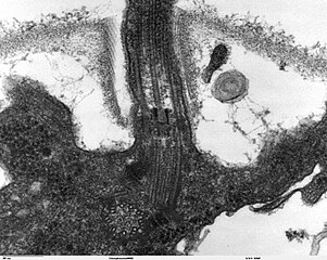
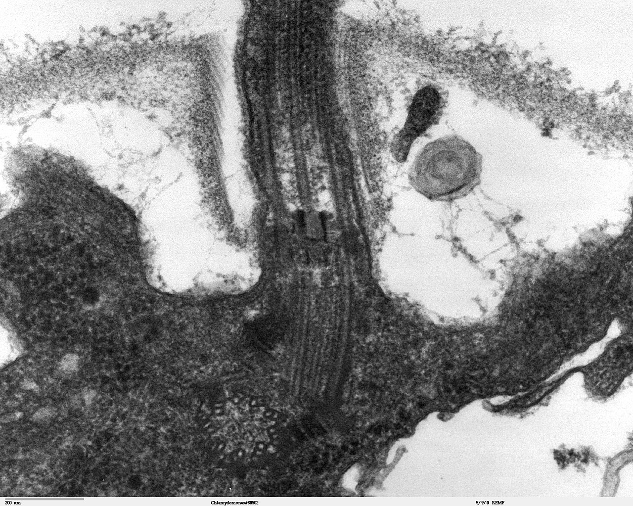
Transmission electron microscope image, showing an example of green algae (Chlorophyta). Chlamydomanas reinhardtii is a unicellular flagellate used as a model system in molecular genetics work and flagellar motility studies. This image is a longitudinal section through the flagella area. In the cell apex is the basal body that is the anchoring site for a flagella. Basal bodies originate from and have a substructure similar to that of centrioles, with nine peripheral microtubule triplets(see structure at bottom center of image). The two inner microtubules of each triplet in a basal body become the two outer doublets in the flagella. This image also shows the transition region, with its fibers of the stellate structure. The top of the image shows the flagella passing through the cell wall. |
| Час створення | |
| Джерело | Source and public domain notice at http://remf.dartmouth.edu/imagesindex.html |
| Автор | Dartmouth Electron Microscope Facility, Dartmouth College |
| Ліцензія (Повторне використання цього файлу) |
Released into the public domain |
| Ця робота була передана у суспільне надбання її автором, Dartmouth Electron Microscope Facility, Dartmouth College. Це застосовується по всьому світу. У деяких країнах це не може бути юридично можливо, в такому випадку: Dartmouth Electron Microscope Facility, Dartmouth College дає кожному право на використання цієї роботи для будь-яких цілей, без будь-яких умов, якщо такі умови не вимагаються за законом.
|
Історія файлу
Клацніть на дату/час, щоб переглянути, як тоді виглядав файл.
| Дата/час | Мініатюра | Розмір об'єкта | Користувач | Коментар | |
|---|---|---|---|---|---|
| поточний | 06:47, 21 вересня 2007 | | 1800 × 1438 (784 КБ) | Neil916 | {{Information |Description= Transmission electron microscope image, showing an example of green algae (Chlorophyta). <br><br>''Chlamydomanas reinhardtii'' is a unicellular flagellate used as a model system in molecular genetics work and flagellar motilit |
Використання файлу
Така сторінка використовує цей файл:
Глобальне використання файлу
Цей файл використовують такі інші вікі:
- Використання в ar.wikipedia.org
- Використання в bs.wikipedia.org
- Використання в ca.wikipedia.org
- Використання в cs.wikipedia.org
- Використання в de.wikipedia.org
- Використання в de.wikibooks.org
- Використання в en.wikipedia.org
- Використання в es.wikipedia.org
- Використання в gl.wikipedia.org
- Використання в id.wikipedia.org
- Використання в ja.wikipedia.org
- Використання в ko.wikipedia.org
- Використання в pl.wikipedia.org
- Використання в ru.wikipedia.org
- Використання в sv.wikipedia.org
- Використання в tr.wikipedia.org
- Використання в zh.wikipedia.org