Смугасте тіло: відмінності між версіями
Kinedw (обговорення | внесок) Створено шляхом перекладу сторінки «Striatum» Мітки: суміш розкладок у тексті Переклад вмісту |
(Немає відмінностей)
|
Версія за 11:36, 23 грудня 2016
| Striatum | |
|---|---|
 purple=caudate and putamen, orange=thalamus | |
 Caudoputamen of the mouse brain | |
| Details | |
| Part of | Basal ganglia[1] Reward system[2][3] |
| Components | Ventral striatum[2][3][4] Dorsal striatum[2][3][4] |
| Identifiers | |
| Latin | neostriatum |
| NeuroLex ID | Striatum |
| TA | A14.1.09.516 A14.1.09.515 |
| FMA | 77616 77618, 77616 |
| Анатомічні назви нейроанатомії[en] | |
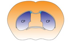
Смугасте тіло (corpus striatum) - підкіркове утворення переднього мозку і надзвичайно важливий компонент екстрапірамідної системи й системи винагороди (англ - Reward System), яке отримує глутамінергічні та дофамінергічні імпульси з різних структур і служить вхідними воротами імпульсів до решти системи базальних гангліїв. У всіх приматів волокнами білої речовини, які називають внутрішньою капсулою, дорзальна частина смугастого тіла ділиться на дві частини, які називаються хвостате ядро і сочевицеподібне ядро.
Вентральна частина складається з прилеглого ядра і нюхового горбка.[4] Смугасте тіло координує декілька когнітивних функцій, включаючи рухи й їхнє планування, прийняття рішень, мотивації, підкріплення й винагороду (Reward System).[2][3][4]
Структура
Типи клітин
Смугасте тіло - гетерогенна структура з точки зору його компонентів - нейронів.[6]
- Шипуваті проекційні нейрони ("середні шипуваті нейрони") являються основними нейронами смугастого тіла.[2] вони є ГАМК-ергічними і, тому класифікуються як гальмівні. Середній шипами проекції нейронів становлять 95% від загальної популяції нейронів у стріатумі людини.[2] Середні шипуваті нейрони мають два основних фенотипи (тобто характерні типи): D1-Тип і D2-тип.[2][4][7]
- Холінергічні інтернейрони вивільняють ацетилхолін, який має цілий ряд важливих ефектів у смугастому тілі. У людини, інших приматів і гризунів, ці інтернейрони реагують на важливі стимули навколишнього середовища стереотипними відповідями, які врівноважуються з відповідями дофамінергічних нейронів чорної субстанції.[8][9] через дофамінові рецептори д5.
- [10]
- Є багато типів ГАМК-ергічних інтернейронів.[6] найбільш відомими є парвальбумін-позитивні, , які беруть участь у потужному прямому гальмуванні основних нейронів.[11] крім того, є ГАМК-ергічні інтернейрони, які тропні до тирозин гідроксилази,[12] соматостатину, синтази оксиду азоту та нейропептіда-Y. [13] [14]
У дорослих людей постійно виробляються нові нейрони в смугастому тілі, і це може зіграти важливу роль у нових методах лікування нейродегенеративних розладів.[15]
Анатомічні структурні елементи


Смугасте тіло поділяється на дорзальну й вентральну частини відповідно до анатомічного розташування і функціональних зв'язків. Вентральний стріатум складається з прилеглого ядра і нюхового горбка, в дорсальний входять хвостате ядро і лушпина.
Дорсальний стріатум може бути диференційований згідно до імунохімічних характеристик — зокрема, по відношенню до ацетилхолінестерази і кaльбіндину (див. мал. "Матриці і стріосомні відсіки").

Вхідні (аферентні зв'язки)
Найважливіші аферентні по кількості аксонів, є кортикостріарні зв'язки. Багато зон кори мозку іннервують дорсальний стріатум. Кіркові пірамідальні нейрони , які віддають свої аксони в стріатум, знаходяться в шарах II-VІ, але найбільш щільні волокна приходять з шару V[17] Вони, в основному, закінчуються на шипах шипуватих нейронів і є глутамін-ергічними нейронами. Інший відомий аферентних шлях - нігростріарний, що починається від нейронів pars compacta чорної субстанції. Також існують аференти від інших елементів базальних гангліїв, таких, як субталамічне ядро (глутамін-ергичні) чи зовнішня (покришкова) частина блідої кулі (ГАМК-ергічні).
Відцентрові (еферентні зв'язки)
Стріарні вихідні (еферентні, відцентрові) шляхи від вентральних і дорзальних компонентів, що складається в основному з середніх шипуватих нейронів проекційних нейронів, які мають два основних фенотипи: "непрямі" з D2-типу рецепторами, і "прямі", з D1-типу рецепторами.[2][4]
Основа базальних гангліїв - це стріатум разом з ділянками, з якими він безпосередньо проекційно пов'язаний через стріато-палідонігральні пучки - щільні, зі слабкомієлінізованими аксонами, що додає їм білуватого забарвлення. Ця проекція складається з покришкової та медулярної частин блідої кулі а також pars compacta і pars reticulata чорної субстанції. Нейрони цієї ділянки гальмуються ГАМК-ергічними синапсами від дорсального стріатума. Серед цих мішеней, бліда куля не посилає аксони за межі системи. Інші посилають аксони до верхніх горбків чотиригорбкового тіла, до додаткової моторної області, до вентральних передніх ядер таламуса, а звідти-в фронтальну ділянку кори.
Функція
Вентральному стріатумі і прилеглому ядрі зокрема, в першу чергу, модерується когнітивна функція системи винагороди (reward cognition), підкріплення, мотиваційної значущості, в той час як су дорзальний стріатум, у першу чергу, регулює пізнавальну рухову функцію, виконавчу функцію, і стимул-відповідь навчання;[2][3][4][18] і лише незначним чином їхні функції перекриваються - у дорсальному стріатумі теж є компонент системи винагороди , що разом із з прилеглим ядром, опосередковує кодування нових рухових програм, пов'язаних з майбутньою нагородою.[3][18]
Метаботропні рецептори дофаміну присутні як на колючих нейронів і на коркових аксонових терминалях[19][20] У людини, смугасте тіло активується стимулами, пов'язаними з системою винагороди, але не тільки - і з неприязню, новизною,[21] несподіванками або іншими інтенсивними подразниками[22] [23]
Клінічне значення
Хвороба Паркінсона
Хвороба Паркінсона призводить до втрати дофамінергічної іннервації в дорсальному стріатумі (та інших базальних гангліїях) та цілого каскаду наслідків. Атрофія стріатума також має місце при хворобі Гентінгтона, хореї, атетозі, і ДЦП.[24]
Залежність
Наркоманія, розлад мозкової системи винагороди, виникає через надекспресія з ΔFosB, а транскрипційний фактор, в D1-Типі середніх шипуватих нейронів в вентральному стріатумі. ΔFosB - ген, який індукується прилеглому ядрі після високих доз наркотиків або інших сильних подразників, що можуть викликати залежність.[25][26]
Біполярний розлад
Генетичні дослідження показали зв'язок між стріарною експресією PDE10A гена й деякими біполярними розладами.[27]
Історія
У XVII і XVIII століттях термін "корпус стріатум" використовується для позначення безлічі різних, глибоких підкіркових елементах.[28] у 1941 році, Сесіль і Оскар Фогт спростили номенклатуру, запропонувавши термін "стріатум" для всіх утворень, побудованих зі стриарних елементів : хвостатого ядра, шлушпини, та інших.
Термін neostriatum був запропонований порівняльними анатомами,які досліджували підкіркові структури у хребетних, і зазначили, що це дійсно філогенетично більш нової частина смугастого тіла. Термін досі використовується в деяких джерелах[29]
Посилання
- ↑ Basal ganglia. BrainInfo. Процитовано 16 August 2015.
- ↑ а б в г д е ж и к л м н Yager LM, Garcia AF, Wunsch AM, Ferguson SM (August 2015). The ins and outs of the striatum: Role in drug addiction. Neuroscience. 301: 529—541. doi:10.1016/j.neuroscience.2015.06.033. PMID 26116518.
[The striatum] receives dopaminergic inputs from the ventral tegmental area (VTA) and the substantia nigra (SNr) and glutamatergic inputs from several areas, including the cortex, hippocampus, amygdala, and thalamus (Swanson, 1982; Phillipson and Griffiths, 1985; Finch, 1996; Groenewegen et al., 1999; Britt et al., 2012). These glutamatergic inputs make contact on the heads of dendritic spines of the striatal GABAergic medium spiny projection neurons (MSNs) whereas dopaminergic inputs synapse onto the spine neck, allowing for an important and complex interaction between these two inputs in modulation of MSN activity ... It should also be noted that there is a small population of neurons in the NAc that coexpress both D1 and D2 receptors, though this is largely restricted to the NAc shell (Bertran- Gonzalez et al., 2008). ... Neurons in the NAc core and NAc shell subdivisions also differ functionally. The NAc core is involved in the processing of conditioned stimuli whereas the NAc shell is more important in the processing of unconditioned stimuli; Classically, these two striatal MSN populations are thought to have opposing effects on basal ganglia output. Activation of the dMSNs causes a net excitation of the thalamus resulting in a positive cortical feedback loop; thereby acting as a ‘go’ signal to initiate behavior. Activation of the iMSNs, however, causes a net inhibition of thalamic activity resulting in a negative cortical feedback loop and therefore serves as a ‘brake’ to inhibit behavior ... there is also mounting evidence that iMSNs play a role in motivation and addiction (Lobo and Nestler, 2011; Grueter et al., 2013). ... Together these data suggest that iMSNs normally act to restrain drug-taking behavior and recruitment of these neurons may in fact be protective against the development of compulsive drug use.
- ↑ а б в г д е ж и к Taylor SB, Lewis CR, Olive MF (February 2013). The neurocircuitry of illicit psychostimulant addiction: acute and chronic effects in humans. Subst. Abuse Rehabil. 4: 29—43. doi:10.2147/SAR.S39684. PMC 3931688. PMID 24648786.
The DS (also referred to as the caudate-putamen in primates) is associated with transitions from goal-directed to habitual drug use, due in part to its role in stimulus–response learning.28,46 As described above, the initial rewarding and reinforcing effects of drugs of abuse are mediated by increases in extracellular DA in the NAc shell, and after continued drug use in the NAc core.47,48 After prolonged drug use, drug-associated cues produce increases in extracellular DA levels in the DS and not in the NAc.49 This lends to the notion that a shift in the relative engagement from the ventral to the dorsal striatum underlies the progression from initial, voluntary drug use to habitual and compulsive drug use.28 In addition to DA, recent evidence indicates that glutamatergic transmission in the DS is important for drug-induced adaptations and plasticity within the DS.50
{{cite journal}}: Обслуговування CS1: Сторінки із непозначеним DOI з безкоштовним доступом (посилання) Помилка цитування: Некоректний тег<ref>; назва «Reward system and psychostimulants» визначена кілька разів з різним вмістом - ↑ а б в г д е ж и к Ferré S, Lluís C, Justinova Z, Quiroz C, Orru M, Navarro G, Canela EI, Franco R, Goldberg SR (June 2010). Adenosine-cannabinoid receptor interactions. Implications for striatal function. Br. J. Pharmacol. 160 (3): 443—453. doi:10.1111/j.1476-5381.2010.00723.x. PMC 2931547. PMID 20590556.
Two classes of MSNs, which are homogeneously distributed in the striatum, can be differentiated by their output connectivity and their expression of dopamine and adenosine receptors and neuropeptides. In the dorsal striatum (mostly represented by the nucleus caudate-putamen), enkephalinergic MSNs connect the striatum with the globus pallidus (lateral globus pallidus) and express the peptide enkephalin and a high density of dopamine D2 and adenosine A2A receptors (they also express adenosine A1 receptors), while dynorphinergic MSNs connect the striatum with the substantia nigra (pars compacta and reticulata) and the entopeduncular nucleus (medial globus pallidus) and express the peptides dynorphin and substance P and dopamine D1 and adenosine A1 but not A2A receptors ... These two different phenotypes of MSN are also present in the ventral striatum (mostly represented by the nucleus accumbens and the olfactory tubercle). However, although they are phenotypically equal to their dorsal counterparts, they have some differences in terms of connectivity. First, not only enkephalinergic but also dynorphinergic MSNs project to the ventral counterpart of the lateral globus pallidus, the ventral pallidum, which, in fact, has characteristics of both the lateral and medial globus pallidus in its afferent and efferent connectivity. In addition to the ventral pallidum, the medial globus pallidus and the substantia nigra-VTA, the ventral striatum sends projections to the extended amygdala, the lateral hypothalamus and the pedunculopontine tegmental nucleus. ... It is also important to mention that a small percentage of MSNs have a mixed phenotype and express both D1 and D2 receptors (Surmeier et al., 1996).
- ↑ Basal ganglia. BrainInfo. Процитовано 16 August 2015.
- ↑ а б Tepper JM, Tecuapetla F, Koós T, Ibáñez-Sandoval O. Front Neuroanat. 2010 Dec 29;4:150. doi: 10.3389/fnana.2010.00150.
- ↑ Nishi A, Kuroiwa M, Shuto T (July 2011). Mechanisms for the modulation of dopamine d(1) receptor signaling in striatal neurons. Front Neuroanat. 5: 43. doi:10.3389/fnana.2011.00043. PMC 3140648. PMID 21811441.
Dopamine plays critical roles in the regulation of psychomotor functions in the brain (Bromberg-Martin et al., 2010; Cools, 2011; Gerfen and Surmeier, 2011). The dopamine receptors are a superfamily of heptahelical G protein-coupled receptors, and are grouped into two categories, D1-like (D1, D5) and D2-like (D2, D3, D4) receptors, based on functional properties to stimulate adenylyl cyclase (AC) via Gs/olf and to inhibit AC via Gi/o, respectively ... It has been demonstrated that D1 receptors form the hetero-oligomer with D2 receptors, and that the D1–D2 receptor hetero-oligomer preferentially couples to Gq/PLC signaling (Rashid et al., 2007a,b). The expression of dopamine D1 and D2 receptors are largely segregated in direct and indirect pathway neurons in the dorsal striatum, respectively (Gerfen et al., 1990; Hersch et al., 1995; Heiman et al., 2008). However, some proportion of medium spiny neurons are known to expresses both D1 and D2 receptors (Hersch et al., 1995). Gene expression analysis using single cell RT-PCR technique estimated that 40% of medium spiny neurons express both D1 and D2 receptor mRNA (Surmeier et al., 1996).
{{cite journal}}: Обслуговування CS1: Сторінки із непозначеним DOI з безкоштовним доступом (посилання) - ↑ Goldberg, JA; Reynolds, JN (December 2011). Spontaneous firing and evoked pauses in the tonically active cholinergic interneurons of the striatum. Neuroscience. 198: 27—43. doi:10.1016/j.neuroscience.2011.08.067. PMID 21925242.
- ↑ Coincident but distinct messages of midbrain dopamine and striatal tonically active neurons. Neuron. 43 (1): 133—43. July 2004. doi:10.1016/j.neuron.2004.06.012. PMID 15233923.
- ↑ Bergson, C; Mrzljak, L; Smiley, J. F.; Pappy, M; Levenson, R; Goldman-Rakic, P. S. (1995). Regional, cellular, and subcellular variations in the distribution of D1 and D5 dopamine receptors in primate brain. The Journal of neuroscience : the official journal of the Society for Neuroscience. 15 (12): 7821—36. PMID 8613722.
- ↑ Inhibitory control of neostriatal projection neurons by GABAergic interneurons. Nat Neurosci. 2 (5): 467—72. May 1999. doi:10.1038/8138. PMID 10321252.
- ↑ Ibáñez-Sandoval, O; Tecuapetla, F; Unal, B; Shah, F; Koós, T; Tepper, JM (2010). Electrophysiological and morphological characteristics and synaptic connectivity of tyrosine hydroxylase-expressing neurons in adult mouse striatum. J Neurosci. 30 (20): 6999—7016. doi:10.1523/JNEUROSCI.5996-09.2010. PMID 20484642.
- ↑ Ibáñez-Sandoval, O; Tecuapetla, F; Unal, B; Shah, F; Koós, T; Tepper, JM (November 2011). A novel functionally distinct subtype of striatal neuropeptide Y interneuron. J Neurosci. 31 (46): 16757—69. doi:10.1523/JNEUROSCI.2628-11.2011. PMID 22090502.
- ↑ English DF, Ibanez-Sandoval O, Stark E, Tecuapetla F, Buzsáki G, Deisseroth K, Tepper JM, Koos T. Nat Neurosci. 2011 Dec 11;15(1):123-30. doi: 10.1038/nn.2984.
- ↑ Neuron-generating brain region could hold promise for neurodegenerative therapies. Science Daily. 20 February 2014. Процитовано 24 February 2014.
- ↑ Reinius B (27 March 2015). Conditional targeting of medium spiny neurons in the striatal matrix.
- ↑ Rosell A, Giménez-Amaya JM (1999). Anatomical re-evaluation of the corticostriatal projections to the caudate nucleus: a retrograde labeling study in the cat. Neurosci Res. 34 (4): 257—69. doi:10.1016/S0168-0102(99)00060-7. PMID 10576548.
- ↑ а б Molecular Neuropharmacology: A Foundation for Clinical Neuroscience (вид. 2nd). New York: McGraw-Hill Medical. 2009. с. 147—148, 367, 376. ISBN 978-0-07-148127-4.
VTA DA neurons play a critical role in motivation, reward-related behavior (Chapter 15), attention, and multiple forms of memory. This organization of the DA system, wide projection from a limited number of cell bodies, permits coordinated responses to potent new rewards. Thus, acting in diverse terminal fields, dopamine confers motivational salience (“wanting”) on the reward itself or associated cues (nucleus accumbens shell region), updates the value placed on different goals in light of this new experience (orbital prefrontal cortex), helps consolidate multiple forms of memory (amygdala and hippocampus), and encodes new motor programs that will facilitate obtaining this reward in the future (nucleus accumbens core region and dorsal striatum). In this example, dopamine modulates the processing of sensorimotor information in diverse neural circuits to maximize the ability of the organism to obtain future rewards. ...
The brain reward circuitry that is targeted by addictive drugs normally mediates the pleasure and strengthening of behaviors associated with natural reinforcers, such as food, water, and sexual contact. Dopamine neurons in the VTA are activated by food and water, and dopamine release in the NAc is stimulated by the presence of natural reinforcers, such as food, water, or a sexual partner. ...
The NAc and VTA are central components of the circuitry underlying reward and memory of reward. As previously mentioned, the activity of dopaminergic neurons in the VTA appears to be linked to reward prediction. The NAc is involved in learning associated with reinforcement and the modulation of motoric responses to stimuli that satisfy internal homeostatic needs. The shell of the NAc appears to be particularly important to initial drug actions within reward circuitry; addictive drugs appear to have a greater effect on dopamine release in the shell than in the core of the NAc. - ↑ Greengard, P (2001). The neurobiology of slow synaptic transmission. Science. 294 (5544): 1024—30. doi:10.1126/science.294.5544.1024. PMID 11691979.
- ↑ Cachope, R; Cheer (2014). Local control of striatal dopamine release. Frontiers in Behavioral Neuroscience. 8: 188. doi:10.3389/fnbeh.2014.00188. PMC 4033078. PMID 24904339.
{{cite journal}}: Обслуговування CS1: Сторінки із непозначеним DOI з безкоштовним доступом (посилання) - ↑ http://www.ucl.ac.uk/news/news-articles/0806/08062502
- ↑ Volman, S. F.; Lammel; Margolis; Kim; Richard; Roitman; Lobo (2013). New insights into the specificity and plasticity of reward and aversion encoding in the mesolimbic system. Journal of Neuroscience. 33 (45): 17569—76. doi:10.1523/JNEUROSCI.3250-13.2013. PMC 3818538. PMID 24198347.
- ↑ Choi EY, Yeo BT, Buckner RL (2012). The organization of the human striatum estimated by intrinsic functional connectivity. Journal of Neurophysiology. 108 (8): 2242—2263. doi:10.1152/jn.00270.2012. PMID 22832566.
- ↑ Walker FO (January 2007). Huntington's disease. Lancet. 369 (9557): 218—28. doi:10.1016/S0140-6736(07)60111-1. PMID 17240289.
- ↑ Nestler EJ (December 2013). Cellular basis of memory for addiction. Dialogues Clin. Neurosci. 15 (4): 431—443. PMC 3898681. PMID 24459410.
- ↑ Olsen CM (Dec 2011). Natural rewards, neuroplasticity, and non-drug addictions. Neuropharmacology. 61 (7): 1109—22. doi:10.1016/j.neuropharm.2011.03.010. PMC 3139704. PMID 21459101.
- ↑ Science Daily: Scientists pinpoint gene variations linked to higher risk of bipolar disorder
- ↑ Raymond Vieussens, 1685
- ↑ MeSH Neostriatum